
Understanding Craniosynostosis: Causes, Symptoms, Complications and Treatment.
Craniosynostosis is a rare defect which affects one in every 2,500-3,000 births, with boys three times more likely to develop the condition. It is characterised by the premature fusion of one or more of the sutures in an infant's skull. The human skull is made up of several bones connected by sutures, allowing for the growth and expansion of the skull as the brain develops. When these sutures close too early, it can lead to abnormal skull shape and potentially impact brain growth. Based on severity, if not treated properly craniosynostosis can lead to number of health issues, involving e.g. developmental delays.
Causes:
The exact cause of craniosynostosis is not always clear, but a combination of genetic and environmental factors is believed to contribute to its development. Genetic mutations may be inherited or occur spontaneously, affecting the normal development of the skull. Environmental factors, such as certain medications taken during pregnancy, maternal smoking, thyroid disease* or advanced paternal age, have also been associated with an increased risk of craniosynostosis.
Symptoms:
The symptoms of craniosynostosis can vary depending on which sutures are affected and how severe the fusion is.
Common signs may include:
- an abnormal head shape,
- a ridge or hard lump along the affected suture,
- an uneven appearance of the face.
Early recognition of the symptoms and checking the baby’s milestones are crucial as premature fusion can cause in some cases complications such as:
- developmental delays caused by increased intracranial pressure,
- cognitive impairment,
- vision problems.
What should I do if I think my child has craniosynostosis:
Craniosynostosis is usually diagnosed through a combination of physical examination, medical history, and imaging scans. A healthcare provider will assess the baby's head shape, feel for abnormal sutures, and inquire about any family history of craniosynostosis. Imaging studies, such as X-rays, CT scans, or MRIs, may be used to visualise the skull and confirm the diagnosis.

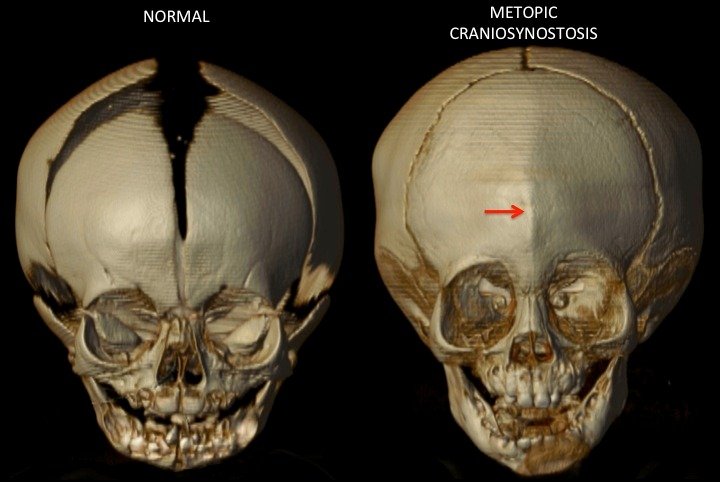
Types of Craniosynostosis:
There are several types of craniosynostosis, depending on which sutures are affected. Some common types include sagittal synostosis, which affects the suture at the top of the skull, and coronal synostosis, which involves the fusion of one of the sutures running from ear to ear.

Treatment:
The primary goal of treating craniosynostosis is to correct the abnormal skull shape and allow for normal brain development. Treatment options vary depending on the severity of the condition and may include:
Surgery: Surgical intervention is often necessary to release the fused sutures and reshape the skull. The timing of surgery depends on the type of craniosynostosis and the age of the child.
Helmets and Orthotic Devices: In some cases, especially with milder forms of craniosynostosis, helmets or orthotic devices may be used to help reshape the skull by applying gentle pressure.
Follow-up Care: Regular follow-up with a healthcare provider is essential to monitor the child's development and address any potential complications, such as increased intracranial pressure.
Pictures:
https://link.springer.com/chapter/10.1007/978-981-19-5458-0_43 Craniosynostosis: A Congenital Anomaly.
https://www.hopkinsmedicine.org/health/conditions-and-diseases/craniosynostosis JOHNS HOPKINS MEDICINE.
https://www.childrens.com/specialties-services/conditions/metopic-synostosis Children`sHealth